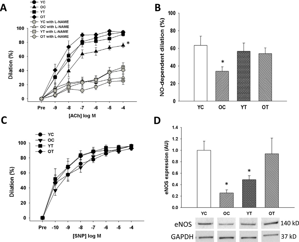
Figure 4. Nitric oxide (NO)-mediated endothelium-dependent dilation.
(A) Endothelium-dependent dilation to acetylcholine (ACh) alone and in the presence of the endothelial NO synthase (eNOS) inhibitor N-G-nitro-L-arginine methyl ester (L-NAME) in young and old control (YC and OC) and young and old TEMPOL-supplemented (YT and OT) mice. (B) NO-dependent dilation (max dilationACh – max dilationACh + L-NAME). (C) Endothelium-independent dilation to sodium nitroprusside (SNP). (D) Aortic protein expression of eNOS expressed relative to GAPDH and normalized to YC mean value. Representative western blot images below. Values are mean ± SEM. (n = 7–8 per group) *p < 0.05 for OC vs. YC, YT and OT.

