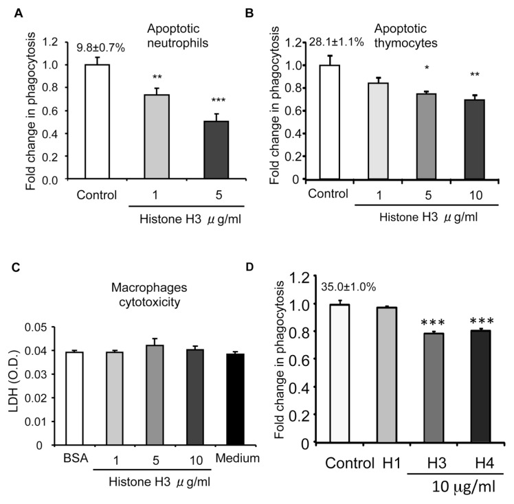
Figure 1.
Histones inhibit efferocytosis. Peritoneal macrophages were incubated with apoptotic neutrophils (A) or apoptotic thymocytes (B) in medium containing 10 μg/mL BSA (control) or histone H3 at increasing concentrations (1, 5 or 10 μg/mL) for 60 min. Efferocytosis assays were then performed as described in Materials and Methods. The percentage of macrophages that phagocytosed apoptotic cells for the control group is shown above the bar. Fold changes were calculated by dividing the percentage of macrophages that phagocytosed apoptotic cells for the experiment groups by that of the control groups. (C) Peritoneal macrophages were exposed to 10 μg/mL BSA (control), histone H3 (1, 5 or 10 μg/mL) or medium for 60 min and then levels of LDH (optical density [O.D.]) in the culture supernatants were determined. (D) Histones H3 and H4, but not histone H1, inhibit efferocytosis. Macrophages were incubated for 60 min with apoptotic thymocytes in media containing BSA (control) or histones H1, H3 or H4 (10 μg/mL), and then efferocytosis assays were performed. *p < 0.05, **p < 0.01, and ***p < 0.001 compared with the control group. The percentage of macrophages that phagocytosed apoptotic cells for the control group is shown above the bar. Fold changes were calculated by dividing the percentage of macrophages that phagocytosed apoptotic cells for the experiment groups by that of the control groups.

