Figure 2.
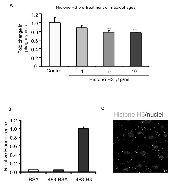
Histone H3 inhibits efferocytosis by binding to macrophages. (A) Macrophages were preincubated with BSA (10 μg/mL) or increasing doses of histone H3 (1, 5 and 10 μg/mL) for 1 h. The macrophages were then washed with fresh medium to remove unbound proteins, and apoptotic thymocytes were added for 60 min, after which efferocytosis assays were performed. (B) Macrophages were incubated with BSA (control), Chromeo 488–conjugated BSA (BSA-488) or Chromeo 488–conjugated histone H3 (H3-488) (5 μg/mL) for 1 h. The cells were then washed three times with PBS to remove unbound proteins. The quantities of proteins bound to macrophages were determined by a fluorescent plate reader. (C) Peritoneal macrophages were plated on coverslips and incubated with 5 μg/mL Chromeo 488–conjugated histone H3 (Histone H3-488) for 1 h. The macrophages were then washed three times with PBS, and confocal fluorescent microscopy analysis was performed to determine bound histone H3. DAPI was used to stain nuclei. **p < 0.01 compared with the control group.

