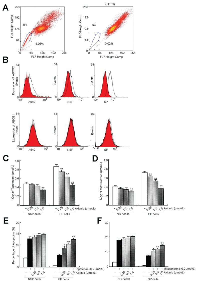
Figure 2.
Axitinib targeted to SP cells and enhanced the efficacy of topotecan and mitoxantrone in the inhibition of proliferation and induction of apoptosis. (A) The A549 cells were stained with Hoechst 33342 as described in Materials and Methods. Gated on forward and side scatter to exclude debris, Hoechst red versus Hoechst blue (R2) was used to sort SP cells. (B) The cell surface expression of ABCG2 and ABCB1. (C, D) Induction of 50% cell death in SP and non-SP cells by topotecan, mitoxantrone and axitinib. Growth inhibition was determined by the MTT assay according to the protocol described in Materials and Methods. (E, F) Sorted SP and non-SP cells treated with toptecan, mitoxantrone and axitinib in the indicated concentrations for 48 h, respectively. Apoptosis was analyzed by flow cytometry as the percentage of cells labeled by annexin V and propidium iodide. All of these experiments were repeated at least thrice, and a representative experiment is shown. Columns, means of triplicate determinations; *P < 0.05; **P < 0.01, compared with topotecan or mitoxantrone treatment.

