
Figure 1. CCR2 is expressed by primary HBMVEC and hCMEC/D3 cells.
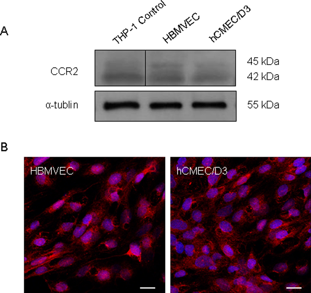
Expression of CCR2, the receptor for CCL2, was evaluated in the brain microvascular endothelial cells used in these studies. (A) 40 µg of total cellular protein from primary human brain microvascular endothelial cells (HBMVEC) and from the HBMVEC cell line hCMEC/D3 were evaluated by Western blot. An equal protein concentration from the THP-1 cell line was used as a positive control for CCR2. Two bands at 45 and 42 kDa represent splice variants of CCR2, CCR2A and CCR2B, respectively. (B) CCR2 expression was also evaluated by immunofluorescence and confocal microscopy. CCR2 (Cy3-red) is expressed in both cell types. Cell nuclei were stained with DAPI (blue). Bar = 18.75 µm.