
Figure 6. PECAM-1 sequesters β-catenin at the membrane in response to CCL2.
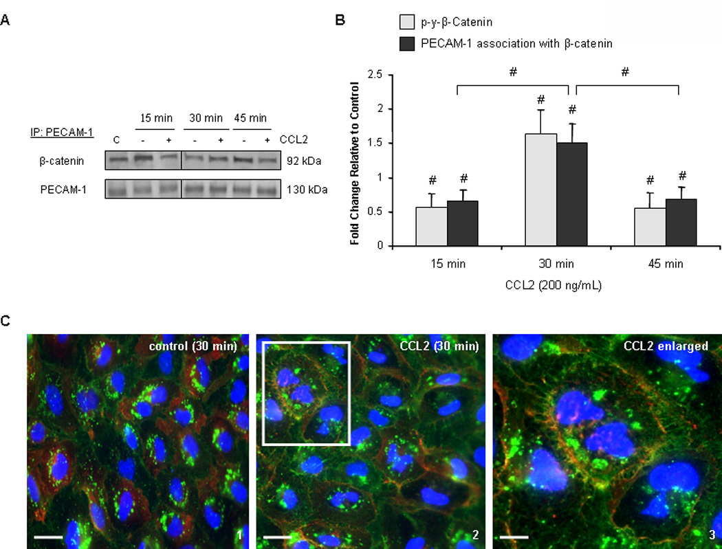
(A–B) 140 µg of total protein were immunoprecipitated from hCMEC/D3 treated with 200 ng/mL CCL2 or diluent. Results were consistent when either PECAM-1 or β-catenin was used in pull-down assay. Blots are representative of 3 independent experiments. An intervening time point was removed from the image and is represented by a dividing line in the blot. Optical densities were compared to OD of immunoprecipitating protein. Densitometry is represented as fold change in CCL2-treated cells relative to control cells. C= control lysate. n=3. (#) p <0.01. β-catenin is found in increased association with PECAM-1 in response to CCL2 relative to control cells and relative to the 15 and 45 minute time points. β-catenin/PECAM-1 association is coincident with β-catenin phosphorylation and dissociation from the adherens junction. (C) Immunocytochemistry was performed on primary HBMVEC treated with 200 ng/mL CCL2 or diluent for 30 minutes. Cells were fixed and permeabilized. β-catenin was stained with Cy3 (red) and PECAM-1 with FITC (green). The third panel is increased magnification of the area outlined in the second panel. After 30 minutes of CCL2 treatment, β-catenin is sequestered at the membrane by PECAM-1.