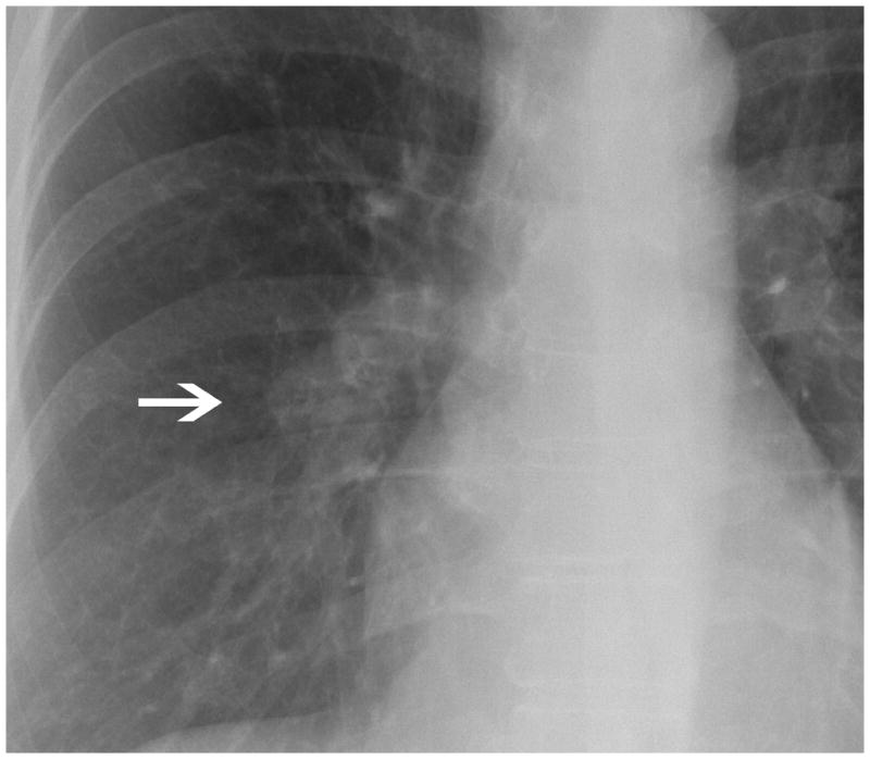
Fig. 1.

There is a poorly defined opacity overlying the inferior aspect of the right hilum (arrow). Some readers considered this abnormality a non calcified nodule while others hilar adenopathy. However, all the readers considered this radiograph highly suspicious for cancer.
