
Figure 1. CCR7 and CCL21 are colocalized on RA synovial tissue (ST) endothelial cells.
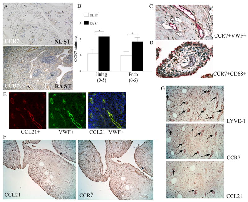
Normal (NL) and and RA ST (A) were stained with anti-human CCR7 (R&D Systems) (original magnification × 200). B. Positive immunostaining was scored on a 0-5 scale and ST lining and endothelial immunostaining are shown as mean ±SEM, n=12. * represents p <0.05. RA synovial tissues were stained for CCR7 (brown staining) and Von willebrand factor (VWF) (red staining) (C) or for CCR7 (brown staining) and CD68 (red staining) (original magnification × 400) (D) in order to distinguish endothelial cells or macrophages that express CCR7. E. Colocalization of CCL21 on VWF+ cells was examined when sections were stained with Texas red labeled anti-goat CCL21 (red) or FITC conjugated anti-rabbit VWF (green staining) or staining overlay (yellow) (original magnification × 400). F. RA serial sections immunostained with anti-CCL21 and anti-CCR7 (original magnification × 200), n=12. G. RA serial sections were immunostained with anti-LYVE-1, anti-CCR7 or anti-CCL21 (original magnification × 400) and CCR7, CCL21 staining were read in 15 LYVE-1+ fields (3 fields in 5 different RA synovial tissues).