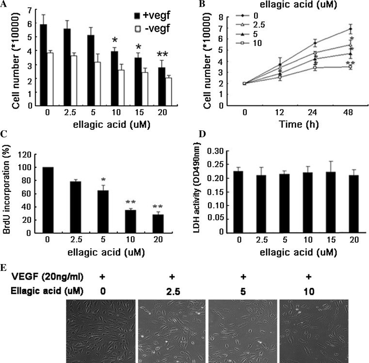
Fig. 1.
Ellagic acid inhibited proliferation of HUVECs induced by VEGF. a After the treatment with ellagic acid at different concentrations ranging from 2.5 to 20 μM for 48 h in the presence or absence of VEGF, cell number was counted. The results showed that the proliferation of HUVECs stimulated by VEGF was significantly decreased in a dose-dependant manner, while ellagic acid had little inhibitory effects on HUVECs that were not stimulated by VEGF (values represent means ± SD, n = 6, *P < 0.05, **P < 0.01, versus untreated control). b Time-course study indicated that ellagic acid could markedly inhibit endothelial cell proliferation in a time-dependant manner (values represent means ± SD, n = 6, *P < 0.05, **P < 0.01, versus untreated control). c Effects of ellagic acid on DNA synthesis were examined by BrdU cell proliferation enzyme-linked immunosorbent assay. The results suggested that ellagic acid could obviously inhibit DNA synthesis of HUVECs in a dose-dependent manner (values represent means ± SD, n = 6, *P < 0.05, **P < 0.01, versus untreated control). d The supernatants of HUVECs after ellagic acid treatment were collected for cytotoxicity examination by LDH cytotoxicity assay kit. The results showed that ellagic acid administration did not result in LDH release from endothelial cells, indicating that ellagic acid posed little cytotoxicity effects upon HUVECs (values represent means ± SD, n = 6). e VEGF-induced HUVECs were treated with ellagic acid at different concentrations for 48 h, and then photographed by at 100-fold magnification for the detection of HUVECs morphological changes. Little abnormal morphological changes of HUVECs were observed after 48-h exposure to ellagic acid. The results further demonstrated that there was no significant toxicity of ellagic acid at any tested concentration in the cellular level

