Fig. 3.
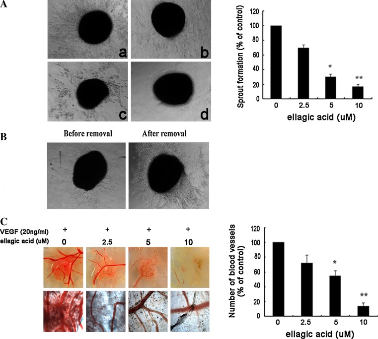
Ellagic acid blocked neo-angiogenesis in chick-associated models. a Ellagic acid dose dependently suppressed sprout formation on the organotypic model of chick aortic ring. Chick aortic rings were embedded in Matrigel and treated with different concentrations of ellagic acid stimulated by VEGF. The changes of sprout formations around various aorta samples were observed on the 3rd day (left panel; a 0 μM, b 2.5 μM, c 5 μM, d 10 μM). The index was defined as a percentage of the untreated control (right panel, values are represented as means ± SD, n = 6, *P < 0.05, **P < 0.01 versus untreated control). b The inhibitory effects of ellagic acid on sprout formation could be reversed after removal of ellagic acid from chick aortic ring. Aortic ring was initially fed with both VEGF and ellagic acid (10 μM) for 48 h (left panel), and then continually treated with VEGF after removal of ellagic acid for an additional 48 h (right panel). Images were representative of three independent experiments. c Ellagic acid could inhibit the microvessels formation on in vivo chick embryonic CAM model. Images were representative of three independent experiments (left panel). The index was defined as the mean number of visible microvessel branch with the defined area of drug-containing pellets on each CAM model (right panel, values are represented as means ± SD, n = 6, *P < 0.05, **P < 0.01 versus untreated control)

