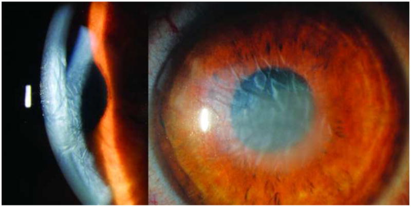
Figure 4.

Slit lamp photo of right and left corneas during the healing process after surgery. The right cornea (left image) shows peripheral graft detachment with corneal clearing in the detached section (superiorly in the image) as compared to the persistent central edema in the central regions of graft adherence, while the left cornea (right image) shows similar peripheral corneal clearing with residual central edema after the Descemet’s stripping only procedure.
