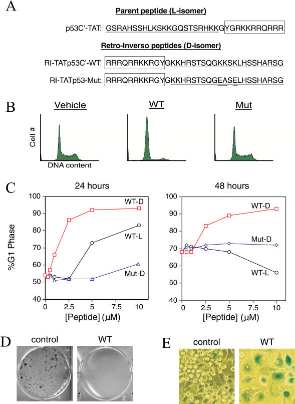
Figure 1. RI-TATp53C′ Induces the Hallmarks of p53 Activity in TA3/St Mammary Carcinoma Cells.
(A) Sequence of p53C′TAT peptide (L-amino acids) and its retro-inverso analogue (D-amino acids). To generate a negative control peptide, three essential lysine residues (Selivanova et al. 1997) were mutated while leaving the remaining peptide sequence intact.
(B) Induction of G1 arrest in TA3/St cells by wild-type RI-TATp53C′, but not mutant peptide, 24 h after peptide addition.
(C) Dose-dependent induction of G1 arrest by RI-TATp53C′ (open square) (D-amino acids) and the less potent p53C′TAT (open circle) (L-amino acids) but not mutant (open triangle) peptide at 24 h (left) and 48 h (right) after single treatment.
(D) Induction of a permanent growth arrest in TA3/St cells by RI-TATp53C′. Cells were treated with RI-TATp53C′ peptide or vehicle for 2 d, replated, and allowed to proliferate in the presence of serum for 10 d. Colonies were then stained with methylene blue.
(E) Induction of a senescence-like phenotype in TA3/St cells by RI-TATp53C′. Cells were treated with RI-TATp53C′ peptide and stained for acidic β-galactosidase activity.

