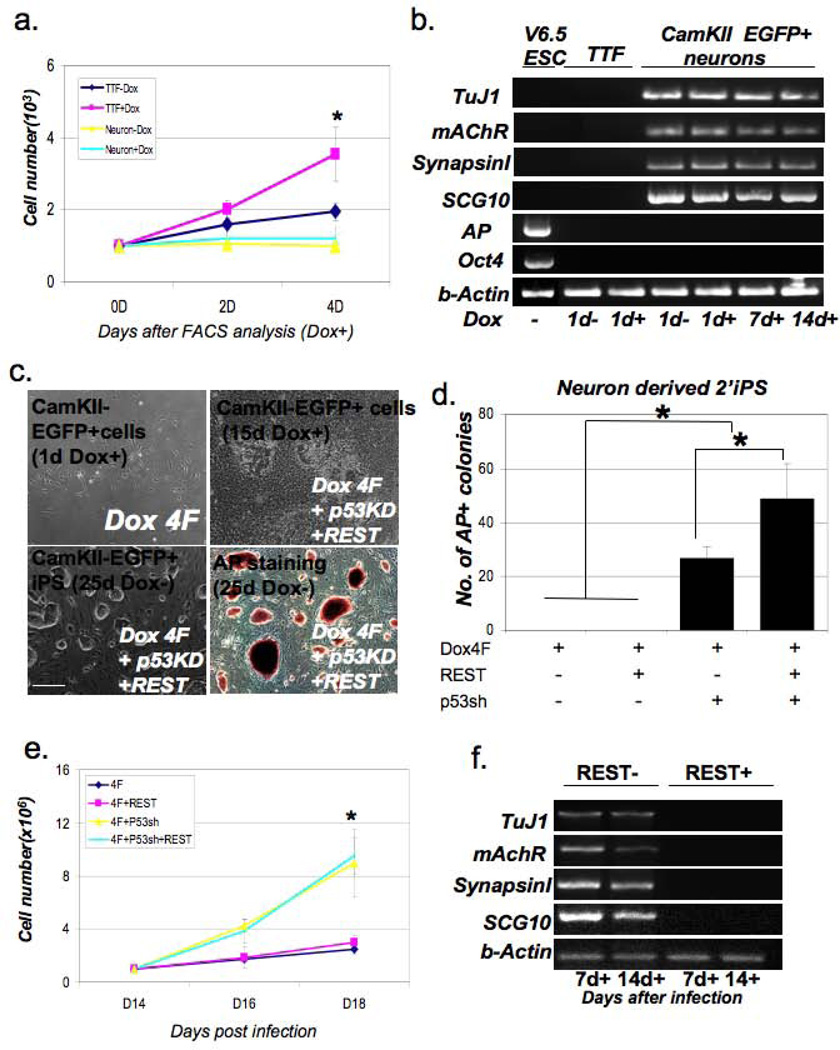
Figure 3. Direct reprogramming of postnatal CamKII-eGFP+ neurons.
(a) Growth curves for secondary fibroblasts and secondary CamKII-eEGFP+ neurons derived from CK-iPS#7 chimeras in the presence or absence of dox. The secondary CamKII-eGFP+ neurons were FACS purified and re-plated after 9 days of dox treatment, whereas secondary fibroblasts derived from the same mice after 9-day dox treatment were dissociated and equal numbers of cells were plated. Cell numbers were counted 2 and 4 days after plating. Data represent mean ± SEM; three independent experiments were performed. ANOVA test, *P < 0.05. (b) Quantitative RT-PCR analysis of neuronal and ES cell specific gene expression during the reprogramming process. (c) FACS purified CamKII-eGFP+ cells were co-infected with a lentivirus expressing a p53shRNA and a lentivirus expressing REST. Cultures were stained for Alkaline Phosphatase activity 3 weeks after infection. Scale bars=200µm (d) The number of Alkaline Phosphatase (AP) positive colonies from FACS-purified CamKII-eGFP+ cells reprogrammed with p53sh and REST 3 weeks after dox treatment. Equal numbers of cells were plated in the presence of doxycycline and the number of AP+ colonies that grew after the withdrawal of doxycyclin was determined 21 days later. 15 individual AP colonies from each group were validated by immunofluorescence staining for Nanog. Data represent mean ± SEM; six independent experiments were performed with three different primary iPS derived eGFP+ cells; ANOVA test, *P < 0.05. (e) Growth curves for CamKII-eGFP+ neurons infected by the additional factors on dox. Equal numbers of FACS purified cells were infected by the additional lentivirial vectors encoding REST and p53sh RNA one day after plating and cultured in the presence of dox. The cell number was determined 2 weeks later. Data represent mean ± SEM; three independent experiments were performed; (f) RT-PCR analysis of neuronal gene expression during reprogramming of eGFP+ neurons by O,K,S,M and p53 shRNA expression in the presence or absence of REST overexpression.

