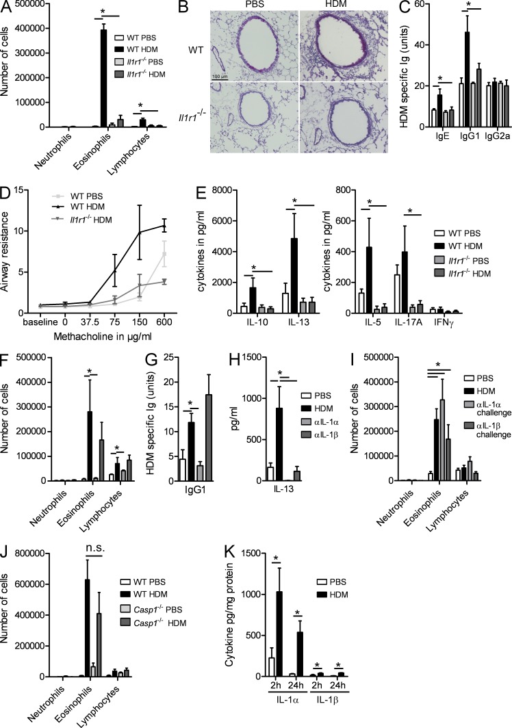
Figure 1.
IL-1RI signaling is crucial for the development of HDM-induced asthma. (A–E) WT and Il1r1−/− mice were sensitized on day 0 with HDM or PBS and were challenged with HDM on days 7–11. (A) Differential cell counts were determined by flow cytometry 72 h later. (B) PAS staining of lung sections. (C) Levels of serum HDM–specific Igs. (D) Airway resistance in response to increasing concentrations of methacholine. (E) Cytokine levels in MLN cells restimulated for 3 d with 15 µg/ml HDM. (F–H) C57BL/6 mice were sensitized with HDM in the presence or absence of blocking antibodies to IL-1α or IL-1β and were challenged with HDM. (F) Differential cell counts were determined by flow cytometry 72 h later. (G) IgG1 levels in sera. (H) IL-13 levels in MLN cells restimulated for 3 d with HDM. (I and J) C57BL/6 mice were sensitized with HDM or PBS and were administered blocking antibodies to IL-1α or IL-1β on the last 3 d of HDM challenge. (I) Differential cell counts were determined by flow cytometry 72 h later. (J) WT and Casp1−/− mice were sensitized with HDM or PBS as a control and were rechallenged with HDM. Differential cell counts were determined by flow cytometry 72 h later. (K) WT mice were administered with PBS or HDM. IL-1α and IL-1β contents were determined in lung homogenates 2 and 24 h later. *, P < 0.05. Results show one representative experiment out of three. Five to six mice/group were used. Results are shown as mean ± SEM.

