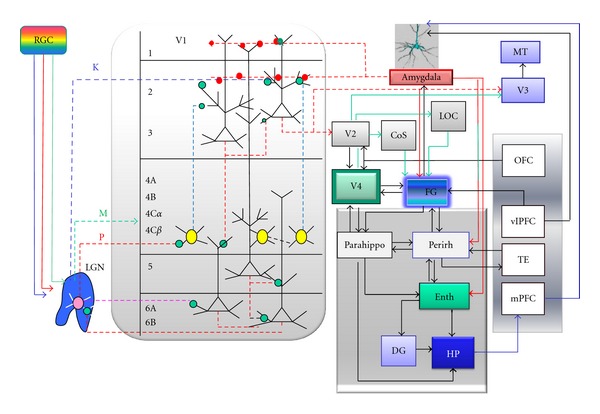
Figure 2.

Visual signal processing along the ventral visual stream. Photons reflected from the object surface traverse first three retinal cell layers to reach photoreceptor-containing cones and rods. Retinal image formation relies mainly on differential glutamate signalling by ON and OFF cones [19, 40]. Local calculations performed by dendritic branches of direction-selective retinal ganglion cells (RGC) and asymmetric nature of synaptic inhibitory inputs from starburst amacrine cells assure high fidelity of object image formation at the retina [41]. Each pixel of the retinal image gets transmitted via dedicated RGC axons to the lateral geniculate nucleus (LGN). Propagating action potentials excite parvocellular LGN neurons (P), which synapse onto stellate cells of V1 (4C layer, yellow) [19]. Direct koniocellular afferents (K) from LGN to L2/3 inform our brain about the relative retinal image displacement (object movement) and activate the dorsal visual stream targeting the orbitofrontal cortex (OFC) [24]. OFC sends rich cholinergic top-down afferents to visual cortex [42, 43] and helps to maintain attention load exercised on V1–V4 areas. The layer L2/3 neurons of V2 send horizontal axonal projections to V4 area, which serve as visual short-term memory buffer [44]. Early visual cortex communicates with fusiform gyrus (FG) and amygdala nuclei of both hemispheres. Primate amygdala projects axons equipped with bouton terminals (dotted red lines and circles) onto dendritic spines located in L1 and L2 layers of V1 [45]. Amygdala-induced neurotransmitter release at the axo-spinous synaptic contacts visual brain areas facilitates formation of visual long-term memories, especially those of high emotional dimension. CoS—collateral sulcus, LOC—lateral occipital cortex, Perirh—perirhinal cortex, Enth—entorhinal cortex, Parahippo—parahippocampal gyrus, DG—dentate gyrus, Hp—hippocampus, vlPFC—ventro-lateral prefrontal cortex, mPFC—medial prefrontal cortex, and TE—temporal lobe.
