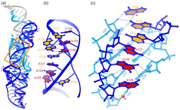
Figure 4.
The oblique A-minor motif forms the interface between the core and the peripheral domains (Klein & Ferré-D’Amaré, 2006). (a) Representation of the entire ribozyme, seen from the direction of P4 and P4.1. In this figure, all the nucleotides in the substrate strand are colored gray. Nucleobases of the ascending and descending strands of the oblique A-minor motif are filled in yellow and red, respectively. Note oblique angle of the region boxed in gold (dark blue) with respect to the core of the ribozyme (cyan). (b) Detailed view of the boxed region in panel (a). Black dashed lines denote hydrogen bonds between nucleobases. (c) Ball-and-stick representation of the oblique A-minor interaction formed between the peripheral domain nucleotides 104–106 and 127–129, and the core domain. Red spheres labeled “w” depict water molecules.

