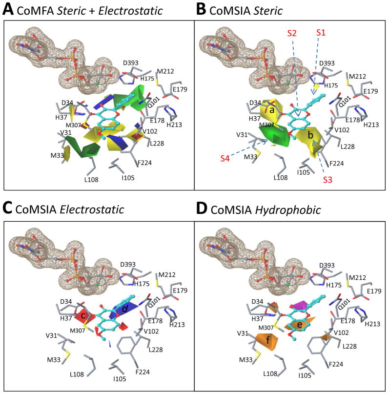
Figure 8. Superposition of the CoMFA/CoMSIA contour maps over the binding site of a homology-modeled UGT1A9 structure based on a simulated binding model of kaempferol (3-OH).
The UGT1A9 protein is shown in a stick model. Kaempferol is indicated in a ball-and-stick model and the cofactor is shown in a ball-and-stick model with a molecular surface. Panel A: Overlay of the CoMFA steric and electrostatic maps with the UGT1A9 binding site. Panel B–D: Overlay of the CoMSIA steric (B), electrostatic (C), and hydrophobic (D) fields with the UGT1A9 binding site.

