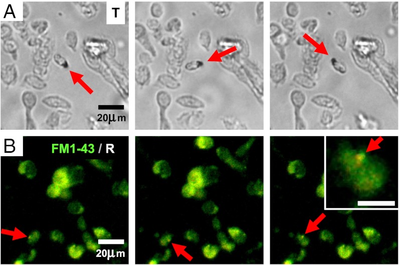
Fig. 1.
Time lapses of cell suspension from dissociated trout olfactory epithelium, showing individual cells rotating with magnetic field. (See Movies S1 and S2 for the two full sequences from which time lapses were extracted). (A) Transmitted light (T), showing an opaque inclusion (red arrow) in the rotating object. (B) Simultaneously recorded dark-field reflection (R) and fluorescence (FM1-43, lipophillic dye), showing reflective objects (white) and cell membrane (green). The rotating cell contains a strongly reflective inclusion (red arrow), displayed as close-up (upper right corner, scale bar 10 μm).

