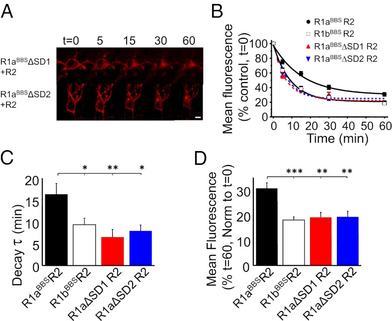
Fig. 5.
SDs in R1a increase the surface stability of R1aR2. (A) Hippocampal neurons expressing either R1aBBSΔSD1R2 or R1aBBSΔSD2R2 with eGFP were incubated in 1 mM d-TC, followed by 3 μg/mL BTX-AF555, for 10 min at RT and imaged at different time points at 30–32 °C. (B) Rates of internalization for BTX-AF555 tagged R1aBBSR2 (●), R1bBBSR2 (□), R1aBBSΔSD1R2 ( ), and R1aBBSΔSD2R2 (
 ) receptors (n = 7–12). (C and D) Exponential decay time constants for membrane fluorescence (C) and extent of constitutive internalization (D) of R1aBBSR2, R1bBBSR2, R1aBBSΔSD1R2, and R1aBBSΔSD2R2. ***P < 0.001; **P < 0.01, *P < 0.05 (one-way ANOVA). (Scale bar: 10 μm.)

