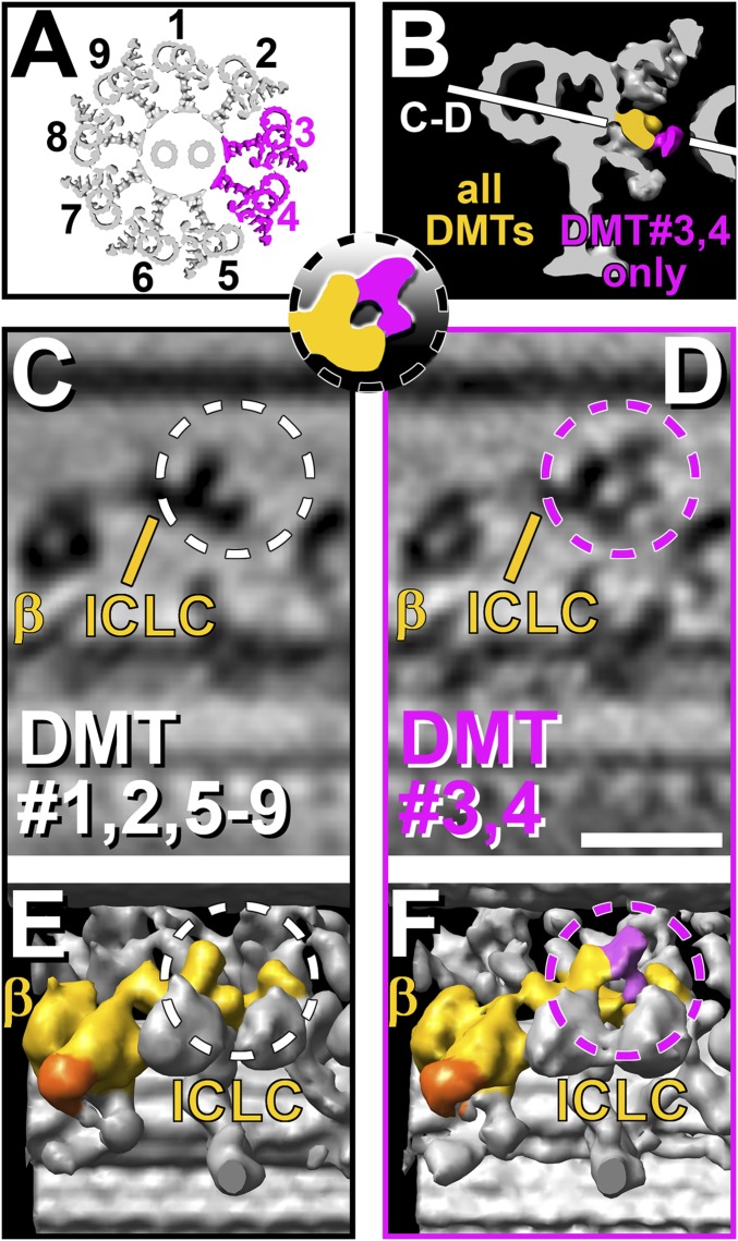
Fig. 3.
Doublet-specific differences of the I1 ICLC structure in pWT axonemes. (A) Simplified schematic model of an axoneme in cross-section highlighting DMTs 3 and 4, which show a doublet-specific feature in the I1 complex, whereas all other DMTs lack this feature. (B and Inset) Isosurface rendering of a DMT in cross-section. The I1 dynein structure present in all DMTs is colored yellow, and the doublet-specific structure present only in DMTs 3 and 4 is highlighted in magenta. Orientation of views shown in C and D is indicated by a white line. (C–F) Tomographic slices (C and D) and isosurface renderings (E and F) of averaged axonemal repeats from pWT viewed from the axoneme center looking outward. Averaged repeats from DMTs 1, 2, and 5–9 (C and E) are compared with averages from DMTs 3 and 4 (D and F). Note the doublet-specific difference: In the average of DMTs 3 and 4, an additional density is present (magenta-colored density and dashed circles in D and F) at the central protrusion, forming a larger ring-shaped structure extending further toward the adjacent B-tubule and the distal protrusion. This additional density is absent from DMTs 1, 2, and 5–9 (white dashed circles in C and E) (Scale bar: D, 20 nm.). Averages of each individual doublet and a second strain, pWT2, are presented in Fig. S4.

