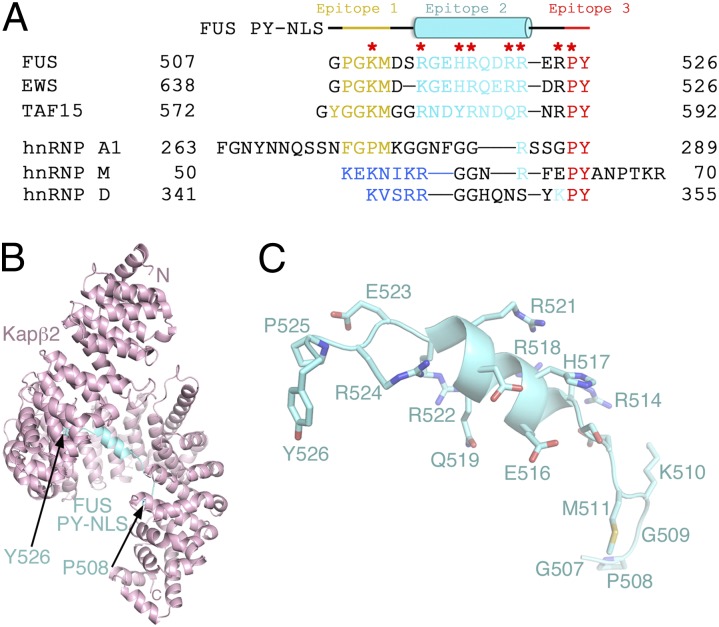
Fig. 1.
The atypical helical PY-NLS of FUS. (A) Sequences of PY-NLSs of FUS, EWS, and TAF15 and hnRNPs A1, M, and D. The N-terminal hydrophobic (yellow) or basic (blue) motifs that form epitope 1 of the NLS, the central epitope 2 (cyan), and the C-terminal epitope 3 (red) are shown. Residues mutated in ALS are marked with red asterisks. (B) Overall structure of the Kapβ2-FUS PY-NLS complex. The karyopherin is pink and the PY-NLS cyan. Side chains of the FUS PY-NLS is shown in C.

