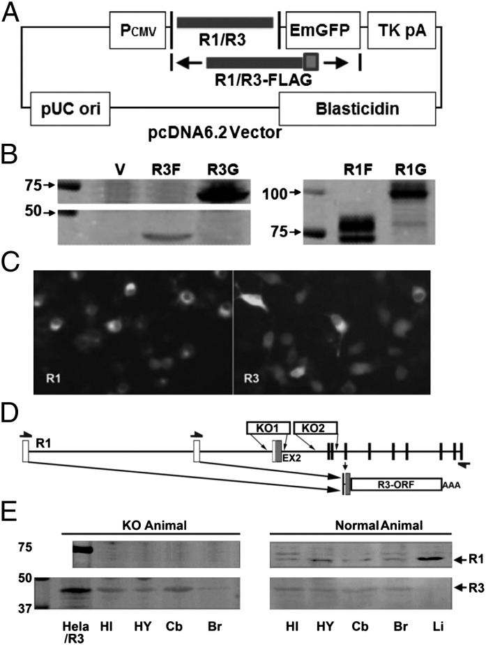
Fig. 2.
IL-1R3 is detected both in vitro and in vivo. (A) Diagrammatic illustration of IL-1R1/R3 cloning into pcDNA6.2 vector. IL-1R1/R3 was inserted after CMV promoter and followed by an in-frame GFP (emerald GFP; EmGFP) protein or a Flag tag. (B) Expression of IL-1R3-Flag (R3F), IL-1R3-GFP (R3G), IL-1R1-Flag (R1F), and IL-1R1-GFP (R1G) detected by Western blot. (C) Immunofluorescence microscopy images of Neuro-2a cells transfected with C terminus GFP-tagged IL-1R1/IL-1R3. (D) Diagrammatic illustration of IL-1R1 KO designs from current IL-1R1 KO lines. The entire IL-1R3 OFR can be detected by RT-PCR in both strains of IL-1R1 KO mice (arrows denote PCR primers). (E) IL-1R3 is detected in various regions of the brain in both IL-1R1 KO and normal animal by Western blot. In contrast, IL-1R1 can only be detected in normal animals. HI, hippocampus; HY, hypothalamus; Cb, cerebellum; Br, brain; Li, Liver.

