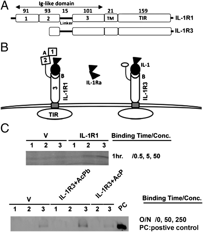
Fig. 3.
IL-1β binds IL-1R3. (A) Diagrammatic illustration of the domains of IL-1R1 and -1R3. TM, transmembrane domain; TIR, tToll/IL-1R domain. Each open box represents a specific domain with numbers indicating the number of amino acids. (B) Diagrammatic illustration of predicted IL-1R3 structure and its potential interaction with IL-1/IL-1ra in comparison with IL-1R1. IL-1R3 is predicted to interact with IL-1 because IL-1R3 retains one (Ig-like domain 3) of the two IL-1 binding sites of IL-1R1. IL-1R3 is unlikely to bind IL-1ra because it does not have the IL-1ra binding site (between the first and the second Ig-like domains of IL-1R1). (C) Detection of cell-bound His-tagged IL-1β with anti-His antibody to IL-1R1–transfected (Upper) or IL-1R3–transfected (Lower) cells. IL-1β binds both receptors specifically [compared with vector (V) group] in a concentration-dependent manner. (Upper) Lanes 1, 2, and 3 correspond to results generated from 0.5, 5, and 50 nM His-IL-1β, respectively. (Lower) Lanes 1, 2, and 3 correspond to results generated from 0, 50, and 250 nM HisIL-1β, respectively.

