Fig. 2.
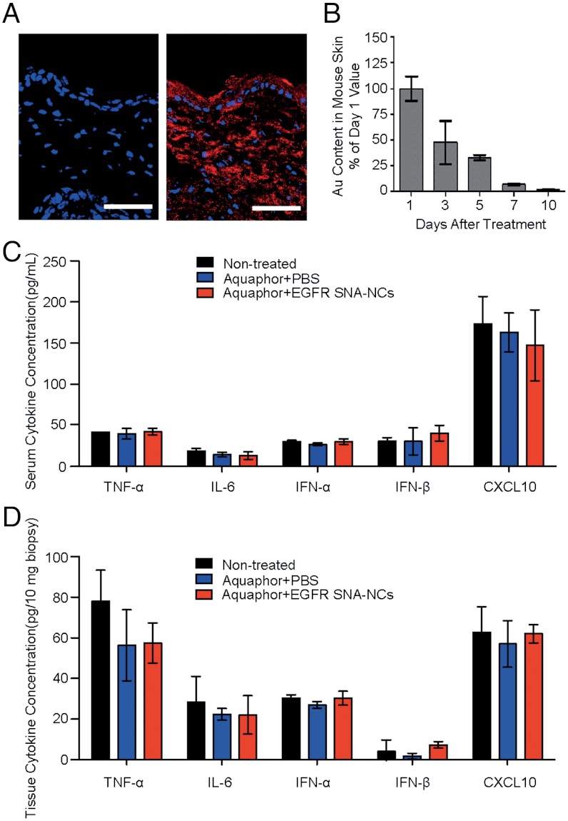
Penetration and clearance of SNA-NCs in hairless mouse skin. (A) Mouse (SKH1-E) skin treated topically with 1∶1 Aquaphor only (Left) or with 50 nM Cy5-labeled (red) SNA-NCs dispersed in the 1∶1 Aquaphor (Right); 3 h after application, the SNA-NCs are seen in the cytoplasm of epidermal cells and the dermis as well. Blue, DAPI-stained nuclei. Scale bars, 100 μm. (B) Mouse skin was treated daily for 3 d with 50 nM nonsense SNA-NCs; samples were harvested at 24 h to 10 d after treatment discontinuation and analyzed by ICP-MS for gold content. The gold content in mouse skin progressively decreases after cessation of topical treatment; 10 d after the final treatment, only 2% of the original gold content remains (n = 3 at each time point). (C) ELISA cytokine assays for TNF-α, IFN-α, IFN-β, IL-6, and CXCL10 in serum show no stimulation of innate immune responses after 3 wk of thrice-weekly treatment (every other day but Sunday; n = 3 per treatment group). (D) EGFR SNA-NC treatment does not induce the cytokine expression locally in the treated skin site after 3 wk of treatment. Results are presented in pg per 10 mg biopsy.

