Figure 1.
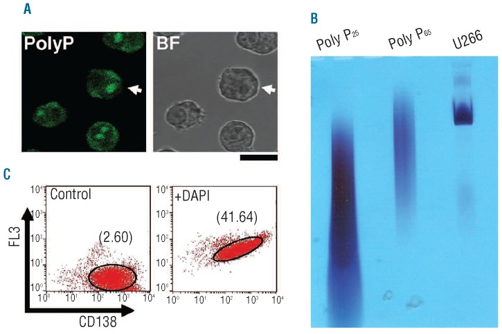
Polyphosphate is present in human myeloma cell lines (HMCL). (A) Localization of cellular polyphosphate (polyP) in the U266 MC line. Fixed cells were treated with DAPI, and specific polyP labeling was determined by confocal fluorescence microscopy (PolyP). The corresponding bright field image is shown (BF). Bar: 10 μm. PolyP cellular distributions in other HMCL (NCI and RPMI) are shown in Online Supplementary Figure S2A. The fluorescence intensity profile of the cell indicated with an arrow is shown in Online Supplementary Figure S2B. (B) Size analysis of polyP from U266 MC. Extracted polyP was separated by 6% urea-polyacrylamide gel electrophoresis (U266) and the gel was stained with toluidine blue to visualize polyP. PolyP25 and polyP65 were used as standards. (C) Flow cytometry analysis of U266 cells labeled with anti-CD138 phycoerythrin antibodies in the absence (Control) and presence of DAPI (+DAPI). The geometric mean fluorescence intensities (MFI) in the FL3 channel of the flow cytometer are indicated. The results of a representative experiment of six are shown.

