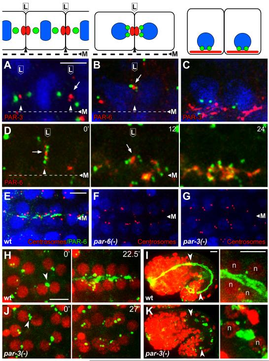
Figure 4.
PAR proteins in centrosome positioning and γ-tubulin localization
(A-C) Intestinal primordial cells in immunostained embryos at (A) metaphase of the E8 to E16 division, (B) shortly after division when paired centrosomes migrate to lateral membranes, and (C) during apical polarization; cartoons of the cells are shown in the upper panels. The images show centrosomes (green; SPD-5 in panel A and IFA1 in, B, C), and PAR-3 or PAR-6 as indicated; nuclei are stained with DAPI (blue). Note that the paired centrosomes in panel B have moved toward the lateral focus of PAR-6. (D) Image sequence from live E16 cells beginning at a stage similar to panel B and showing centrosomes (green, γ-tubulin:GFP) and PAR-6 (red, PAR-6:Cherry); time in minutes at upper right. Note that the centrosome pairs in both cells move apically with the focus of PAR-6, and that PAR-6 spreads after reaching the apical surface. (E-G) Images of the E16 primordium in immunostained wild-type embryos and embryos depleted of both maternal and zygotic PAR-6 or PAR-3 (n=25 and 27 embryos, respectively). Note that most of the paired centrosomes (red, IFA) are apical after depletion of PAR-6 but not after depletion of PAR-3. (H-K) γ-tubulin (green, γ-tubulin:GFP) and nuclei (red, histone:Cherry, ‘n’) in wild type embryos (H,I) and embryos depleted of PAR-3 (J,K). Note the failure of centrosomes and γ-tubulin to localize apically in the PAR-3-depleted embryo. The left panels in I and K show low magnifications of entire embryos during later morphogenesis, with the intestines indicated by arrowheads; the right panels show high magnification views of some of the intestinal cells. γ-tubulin is apical in the wild-type embryo, but ectopic in the PAR-3-depleted embryo. Scale bar (A-C, 2.5μm, E-K, 5μm). See also Figure S4, Movie S4, and Movie S5.

