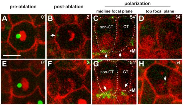
Figure 6.
Centrosomes are required for accumulation of γ-tubulin at the apical surface
Live images of two wild type embryos (A-D and E-H) before (t=0) and after (t=2 minutes) laser ablation of the centrosome, and again after 54 minutes; reporters as in Figure 1. γ-tubulin normally accumulates at the midline before 1 hour, as is visible in the non-centrosome ablated (non-CT) cells, but has not accumulated in the cells with the ablated centrosomes (CT). Note that the nuclei in CT cells have not localized to the midline, but are instead at different focal planes (top focal plane, D and H). The arrow in panel H indicates a non-apical, small cytoplasmic focus of γ-tubulin that likely corresponds to a fragment of the ablated centrosome. CT cells have levels of cytoplasmic γ-tubulin that are comparable to non-CT cells, but lack apical accumulation; the apical surface of CT cells showed a statistically insignificant enrichment of γ-tubulin (−.0018±.012, n=19) compared to non-CT cells (.2081±.082, n=38, two tailed t-test; p ≤ 1.83×10⁻¹⁷). Scale bar = 5μm. See also Movie S7.

