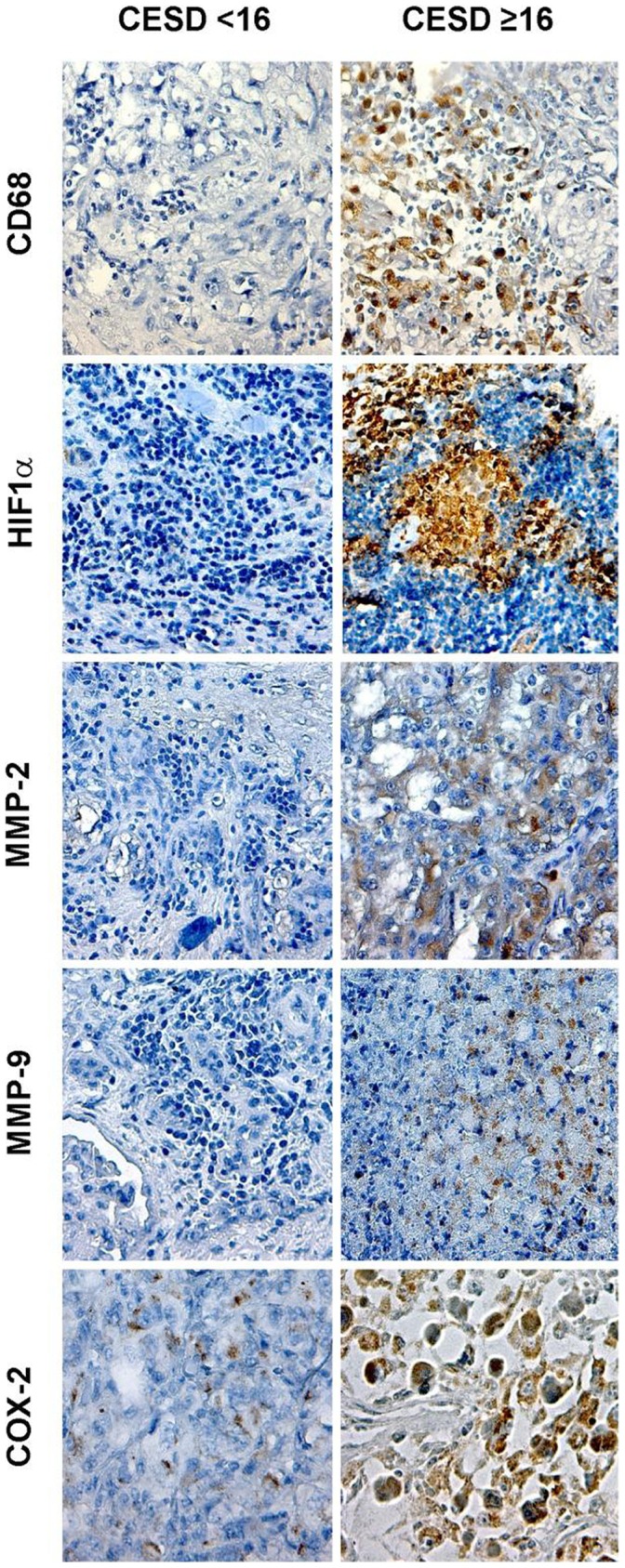
Figure 3. This figure shows representative images of immunohistochemical staining for CD68, HIF1α, MMP-2, MMP-9, and COX-2 in patients scoring greater or equal to 16 versus less than 16 on the CES-D.

Pictures were taken at original magnification X200.
