Figure 2.

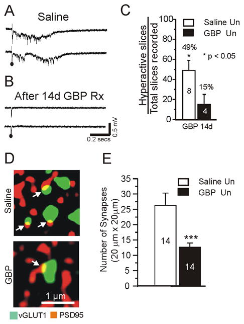
Chronic treatment with gabapentin (GBP) decreases evoked epileptiform discharges and excitatory synapse density in undercut cortex.
A: Representative field potential responses from layer V in undercut region of in vitro slice. Recordings from saline-treated rat 14 d after lesion placed. Just suprathreshold extracellular stimuli evoked epileptiform potentials. B: Same protocol as A, except rat had sc GBP infusion (10 mg/kg/d) × 14d following lesion. Recordings made on day 14 of GBP infusion. Stimuli failed to evoke epileptiform responses. C: Incidence of evoked epileptiform activity in undercut cortical slices in GBP and saline treated animals. Numbers in bars: number of animals. Average of 4.3 slices was assessed per animal. Data are expressed as percentage of slices with evoked epileptiform activity. *: p < 0.05. White bars: saline controls; black bars: GBP. D–E: Chronic GBP treatment (10 mg/kg/d sc × 7d) decreases density of excitatory synapses in undercut cortex. Data obtained from animals on 7th day of treatment following undercut.. D: Confocal images from sections of saline-treated and GBP-treated undercut rats. Double immunocytochemistry for vGlut1 (green, 1:500) and PSD95 (red, 1:200). Arrows: Sites of close apposition and presumptive synapses (yellow). E: Quantification of co-localization of pre and postsynaptic markers vGlut1 and PSD95 in undercut cortical sections from saline-treated (white bar: n =14 sections from 5 animals) and GBP-treated animals (black bar: n = 14 sections from 5 animals with 2–3 sections/rat). Three confocal images taken from each section. ***:p < 0.001. Data are expressed as mean ± SEM. Un: undercut.
