Figure 2.

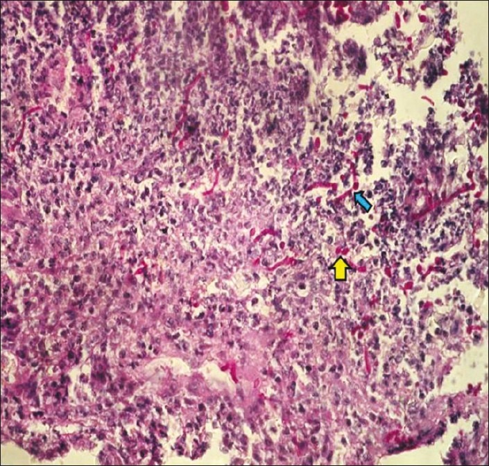
Photomicrograph showing granulation tissue, numerous yeast (yellow arrow), and pseudohyphal fungal forms (block blue arrow) on Periodic acid-Schiff stain consistent with Candida albicans

Photomicrograph showing granulation tissue, numerous yeast (yellow arrow), and pseudohyphal fungal forms (block blue arrow) on Periodic acid-Schiff stain consistent with Candida albicans