
Figure 3.
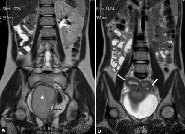
(a) Coronal Single shot T2W image shows absence of the right kidney. The distended hemivagina (asterisk) is seen on the right side and the normal collapsed left hemivagina with minimal fluid is seen adjacent to it (black arrow). The distended hemivagina ends above the introitus and its contents are hypointense to fat. (b) Coronal Single shot T2W image shows right and left uterine horns (white arrows). The right uterine horn cavity is seen to communicate with the upper end of the fluid collection in right hemivagina (small black arrow)