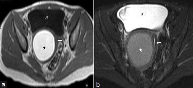
Figure 4.

(a) Axial T1W image of the pelvis shows bright signal intensity of the right hemivaginal collection (asterisk). The collapsed left hemivagina is seen adjacent to it (arrow) (b) Axial fat saturated T2W image of the pelvis showing right hemivaginal collection (asterisk) and collapsed left hemivagina with minimal fluid (arrow)
