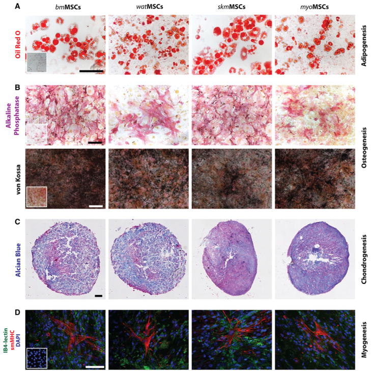
Fig. 3.
Multilineage differentiation of MSCs. a Differentiation into adipocytes was revealed by oil red O staining (scale bar 100 μm). b Differentiation into osteocytes was revealed by alkaline phosphatase staining as well as von Kossa staining for calcium mineralization (scale bar 100 μm). Insets represent MSCs in non-differentiating control medium. c Chondrogenic differentiation was revealed in pellet culture by the presence of glycosaminoglycans, detected by Alcian blue staining (scale bar 200 μm). d Smooth muscle cell (SMC) differentiation was evaluated by culturing MSCs in the presence of murine mDECs for 7 days. Induction of SMC phenotype was assessed by the expression of smooth muscle myosin heavy chain (smMHC). IB4-FITC lectin was used to stain mDECs and DAPI for cell nuclei (scale bar 100 μm). Inset represents MSCs cultured in the absence of mDECs

