Fig. 6.
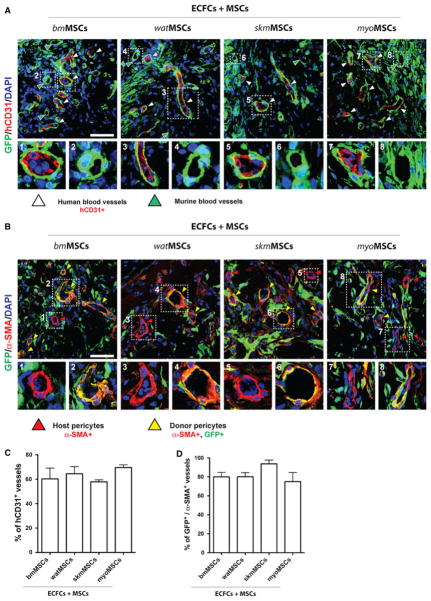
Perivascular engraftment of MSCs. GFP-MSCs obtained from four different murine tissues were combined with ECFCs (2 × 10⁶ total; 40:60 ECFC/GFP-MSC ratio) in 200 μL of Matrigel and the mixture subcutaneously injected into nu/nu mice (n = 4 for each MSC group). a Immunofluorescent staining of engrafted MSCs and ECFCs using anti-GFP and anti-human CD31 antibodies, respectively. White arrowheads indicate hCD31+, ECFC-lined vessels. High magnification of selected regions showing representative human (panels 1, 3, 5, 7) and mouse (panels 2, 4, 6, 8) blood vessels that were covered by perivascular GFP-MSCs. b Immunofluorescent staining of engrafted MSCs and perivascular cells using anti-GFP and anti-α-SMA antibodies, respectively. α-SMA-expressing perivascular cells were either donor GFP+ MSCs (yellow arrowheads) or GFP− host cells (red arrowheads). High magnification of selected regions showing representative blood vessels with GFP+ donor (panels 2, 4, 6, 8) and GFP− host (panels 1, 3, 5, 7) perivascular cells. All images are representative of explants from four different mice. Nuclei were counterstained with DAPI. (scale bars 50 μm). c Percentage of blood vessels that were lined by hCD31+ ECFCs. d Percentage of blood vessels with perivascular cells that expressed both GFP and α-SMA. Bars represent mean values determined from four replicate implants ± SEM

