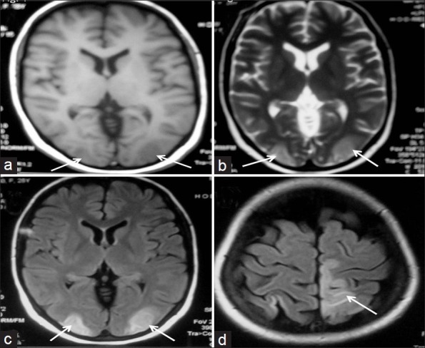
Figure 1.

MR images showing hypointensities on TIW (a) and hyperintensities on T2W (b) in bilateral occipital lobes. FLAIR images (c, d) showing hyperintensities in the bilateral occipital lobes and left high parietal region with sparing of the calcarine and paramedian occipital lobe structures
