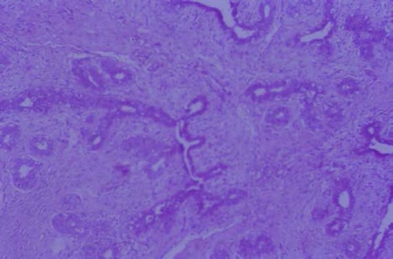
Figure 3a.

Sections of the previously shown breast mass demonstrating proliferating glands and stroma with intracanalicular and pericanalicular patterns of growth, dilated spaces and leaf like projection. Glands show focal apocrine metaplasia and mild focal epithelial hyperplasia.
