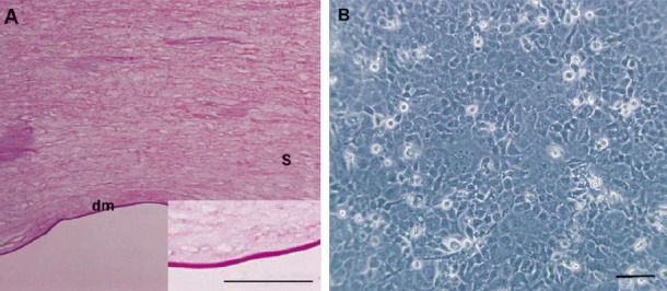
Fig. 1.

Microscopic images of APCM and B4G12 cells. (A) HE staining showed that the acellular porcine corneal matrix was cell-free and the collagen structure was preserved. A high magnification image of APCM is shown in the lower right corner. (B) Monolayer of B4G12 cells formed after 72 h of sub-cultivation. Scale bars: A=50 μm, B=100 μm. dm= Descemet's membrane; s= stroma.
