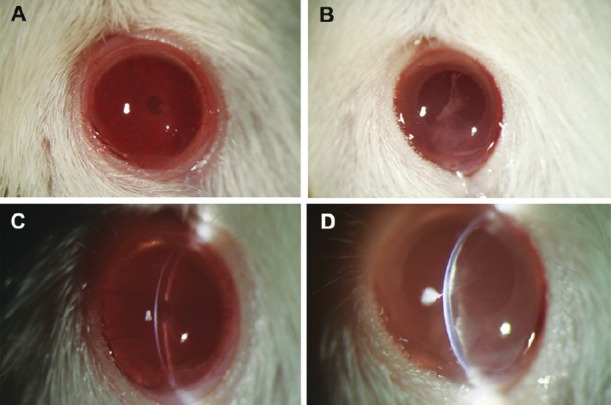
Fig. 2.

Representative corneal photos after the surgery. (A) Cornea in the B4G12 cell-injection group was clear, the anterior chamber and the iris were clearly visible. (B) In contrast, cornea in the cryoinjury only group was opaque. (C) The slit image of the cornea in the B4G12 cell-injection group showed that the cornea was at normal thickness, and the endothelium surface was smooth. (D) The slit image of the cornea in the cryoinjury only group showed that the cornea was swollen and there was lamellar keratic precipitate on Descemet's membrane.
