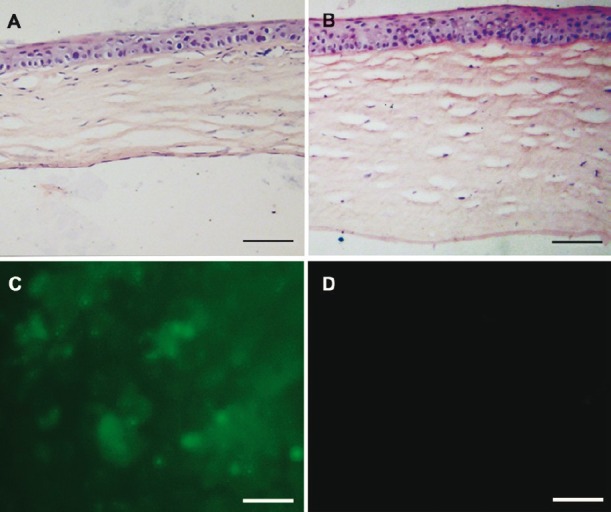
Fig. 3.

Histological examination of the corneas after the surgery. (A) HE-stained cross-section showed that Descemet's membrane was covered by cell monolayer in the B4G12 cell-injection group. (B) No cells were present on Descemet's membrane in the cryoinjury only group. Note that the corneal stroma was much thicker than that in the B4G12 cell-injection group. (C) Fluorescence microscopy confirmed that cells covering the Descemet's membrane in the B4G12 cell-injection group were CFDA SE-positive. (D) No signal was detected from the corneas of the cryoinjury only group. Scale bars: 100 μm.
