
Fig. 4.
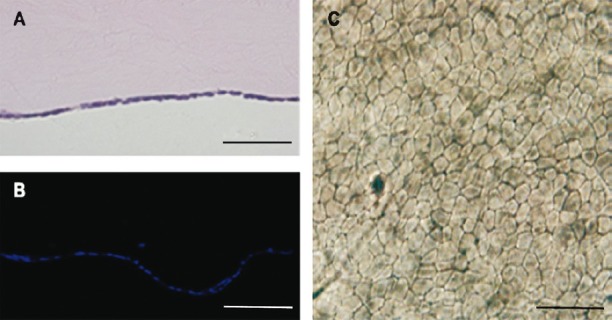
Histological analysis of the corneal endothelium equivalents. (A) HE staining showed there was a monolayer of cells covering the Descemet's membrane of the APCM. (B) DAPI staining confirmed the cell coverage of the Descemet's membrane of the APCM. (C) Trypan blue and alizarin red S co-staining showed the B4G12 cells were alive and clearly delineated the cell boundaries. Scale bars: 50 μm.