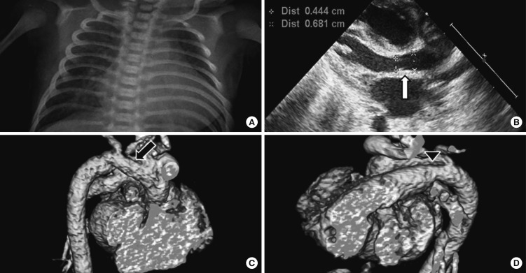
Fig. 2.
Data from echocardiography and computed tomography examinations. (A) Cardiomegaly on chest X-ray. (B) Mild hypoplasia of aortic valve through the echocardiography (arrow). (C) Hypoplasia of arotic arch through the 3-dimensional cardiac computed tomography (arrow). (D) Large patent ductus arteriosus through the three-dimensional cardiac computed tomography (arrowhead).

