Figure 1.

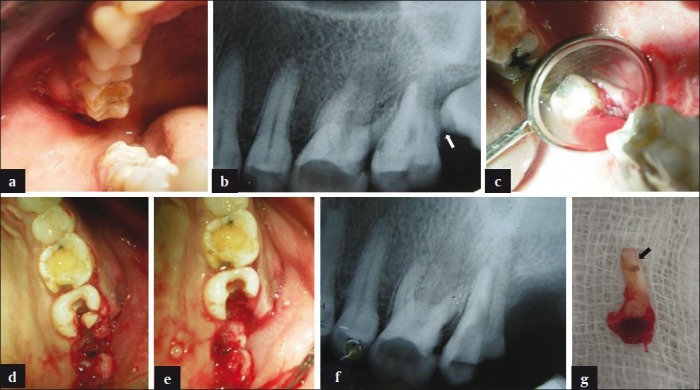
(a) Clinical view of intraoral swelling in relation to 17 and 18. (b) Radiographic view showing resorption in distobuccal root of 17 (white arrow) due to impingement of 18. (c) Surgical view, following extraction of 18, revealing bone loss around distobuccal root of 17. (d) Clinical view of 17 following vertical cuts and separation of its distobuccal root. (e) Surgical view following extraction of 18 and resection of distobuccal root of 17. (f) Radiographic view following resection and extraction of distobuccal root of 17. (g) Extracted distobuccal root of 17 showing resorption (black arrow)
