Figure 1.
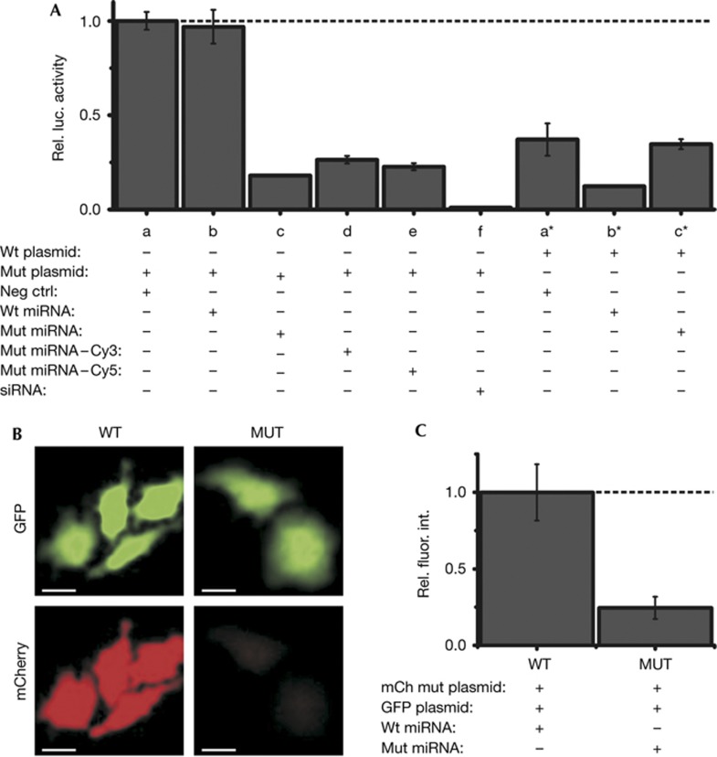
Effect of fluorophore modification and microinjection on miRNA function. (A) Luciferase reporter assays of HeLa cells co-transfected with luciferase reporter plasmids bearing the wild-type (wt) or a mutant (mut) 3′ untranslated region of mouse HMGA2, and either a negative control siRNA (neg ctrl), wild-type let-7a-1 (wt miRNA) or mutant let-7a-1 (mut miRNA) miRNA. An siRNA, Siluc2, was used as a positive control for repression. Renilla luciferase activity was used for internal normalization of firefly luciferase activity within each sample. All samples were normalized with respect to negative control (a). Results presented are from four replicates. Error bars, standard deviations. (B) Representative images of GFP fluorescence (top) and mCherry fluorescence (bottom) in cells coinjected with an mCherry reporter plasmid, a GFP control plasmid and, either the wild-type (WT) or mutant let-7a-1 (MUT) miRNA are shown. Scale bar, 20 μm. (C) Quantification of mCherry fluorescence relative to GFP fluorescence from B, normalized with respect to the WT sample (n=3 independent trials, 50 cells per group). Error bars, s.e.m. GFP, green fluorescent protein; miRNA, microRNA; siRNA, small interfering RNA.

