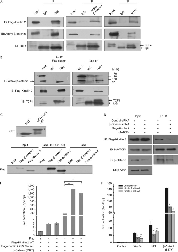
Figure 3.
Kindlin 2 forms a complex with active β-catenin and TCF4 to enhance Wnt signalling. (A) Nuclear extracts were prepared from MCF7–Flag–Kindlin 2 stable cells and co-immunoprecipitation (Co-IP) was performed. (B) Total lysates were extracted from MCF7–Flag–Kindlin 2 stable cells for two-step Co-IP. The first IP was performed with FLAG-M2 beads or mouse immunoglobin (Ig)G. The eluate from the first IP was then used for the second IP with anti-TCF4 antibody or mouse IgG. The immunoprecipitates from each step were detected by western blot (WB). (C) Glutathione S-transferase (GST) or GST–TCF4 1–53 was evaluated by WB (upper panel). Flag, Flag–β-catenin, or Flag–Kindlin 2 were separately transfected into 293T cells. Total protein was extracted and incubated with GST or GST–TCF4 for GST pulldown. (D) Flag–Kindlin 2 and HA–TCF4 were co-transfected into 293T cells, in the presence of control or β-catenin siRNA. Protein was extracted for co-IP. (E) Cells (293T) were transfected as the indicated plasmids plus the SuperTop/Fopflash plasmids for 24 h. (F) After 24 h transfection with control or Kindlin 2 siRNAs, 293T cells were transfected again with SuperTop/Fopflash for 24 h and then treated with Wnt3a (200 ng, 6 h), LiCl (20 mM, 6 h), or 293T cells were co-transfected again with β-catenin (S37Y) and SuperTop/Fopflash for 24 h. Error bars indicate s.d. values, n=3; *indicates P<0.05 by Student's t-test. HA, haemagglutinin; LiCl, lithium chloride; siRNA, small interfering RNA; TCF4, T-cell factor 4; WT, wild-type.

