Figure 4.
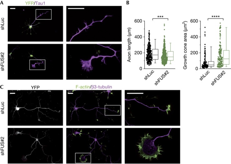
FUS knockdown affects axon and growth cone morphology. Hippocampal neurons co-transfected with the shLuc or shFUS#2 and pEYFP-C1 before plating. (A) Immunostaining at day 4 with anti-YFP as transfection control and anti-Tau1 as axonal marker. Right panels show a high-magnification view of growth cones. For morphometric analysis processes with proximal-to-distal Tau1 gradient were defined as axons. (B) Quantification of axonal length (left, n=325 for shLuc, n=388 for shFUS#2) and growth cone area (right, n=147 for shLuc, n=164 for shFUS#2) measured blinded to the experimental condition. Box-plots represent lower quartile, median and upper quartile. Whiskers represent the 5th and 95th percentile. Mann–Whitney test: ***P<0.001, ****P<0.0001. (C) Staining for F-actin (using phalloidin) and β3-tubulin. Right panels show a high-magnification view of axonal growth cones. Scale bars, 25 μm. FUS, Fused in sarcoma; shFUS, short hairpin FUS; shLuc, short hairpin RNA targeting the luciferase transcript; YFP, yellow fluorescent protein.

