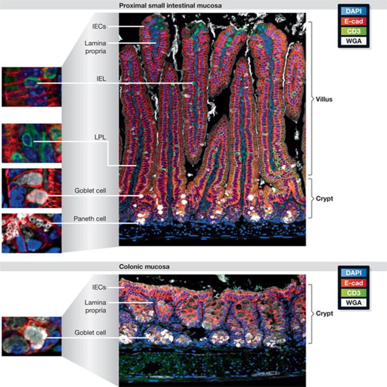
Figure 1.
Structural features of the intestinal mucosa. Paraffin-embedded, formalin-fixed sections of small intestinal (upper panel) and colon tissue (lower panel) from a C57Bl/6 mouse were stained as described elsewhere [49]. E-cadherin (E-cad) is shown in red and CD3 in green. The highly polarized epithelium of the small intestine is organized in a crypt–villus structure. Crypts provide a protective niche for proliferating stem cells (not depicted), and continuous migration along the crypt–villi axis is accompanied by IEC differentiation. The Paneth cells (small white vesicles) are next to the stem cells at the bottom of the crypts, and goblet cells (large white vesicles) are dispersed along the crypt and lower villus region. These two cell types generate the mucus barrier. T cells are shown as an example of intestinal immune cells; lymphocytes can be seen in between or adjacent to the epithelium (IELs) or within the lamina propria (LPL). Epithelium organization in the colon: the crypts end in a flat surface without villi, generating a smoother mucosal tissue. A large number of goblet cells produce a dense mucus layer that covers the epithelium (not depicted). There are no Paneth cells in the healthy colon and the number of immune cells in the lamina propria is much lower than in the small intestine. IEC, intestinal epithelial cell; IEL, intraepithelial lymphocytes; LPL, lamina propria lymphocytes, WGA, wheat germ agglutinin.

