Figure 3.
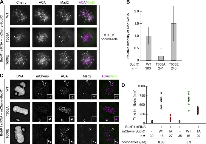
BubR1 autophosphorylation is required for efficient kinetochore targeting of Mad2 and a prolonged mitosis induced by nocodazole. (A and C) Kinetochore localization of mCherry-BubR1 (mCherry; by an anti-mCherry antibody), ACA, and Mad2 in cells expressing BubR1 siRNA and siRNA-resistant mCherry-tagged wild-type (WT) BubR1 or BubR1 phosphorylation mutants as indicated and treated with 3.3 µM nocodazole (A) or not treated (C). In C, a pair of sister kinetochores on unaligned chromosomes indicated by arrows is shown in the insets. Bars: (main images) 10 µm; (insets) 1 µm. (B) Quantitation of the normalized integrated intensity of Mad2 signals against ACA signals at kinetochores in cells treated with 3.3 µM nocodazole. The experiments were repeated twice, and the numbers of kinetochores from ≥10 different cells quantified for each condition were shown. Error bars represent standard error (*, P < 0.05). (D) Time spent in mitosis (measured as the time of a cell’s rounding up by time-lapse imaging) at the indicated nocodazole concentrations for H2B-EYFP cells transfected with BubR1 siRNA along with or without an mCherry expression vector fused with wild-type BubR1 (WT) or the nonphosphorylatable BubR1 mutant (TA) as indicated. Each dot represents a single cell, and the numbers of cells filmed are presented. We should note that a majority of the cells expressing wild-type BubR1 were still in mitosis when the filming was ended; therefore, the time in mitosis for those cells showed here is underrepresented.

