
Figure 4. Arginine 69 Is Important for CpdR-Mediated PdeA Degradation.
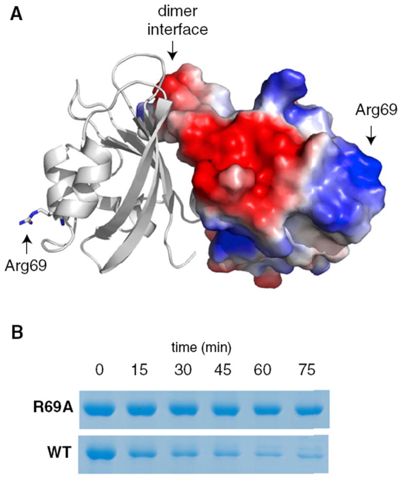
(A) Cartoon/surface representation of PAS dimer with right hand subunit surface colored by electrostatic potential (blue: positive; red: negative) using the qualitative vacuum electrostatic potential display native to Pymol (Schrödinger, LLC). Arginine 69 is modeled in stick representation on the left hand subunit and its location is marked with arrows on both subunits. See Figure S3 for the electron density map of the region surrounding this residue.
(B) Mutation of Arg69 to alanine (R69A) substantially inhibits PdeA degradation by CpdR/ClpXP, likely due to its failure to interact with CpdR (Figure S4).