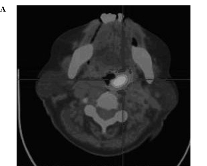
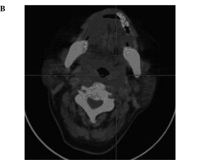
Figure 1.


(A) PET-CT image of a patient (male, NHL DLBCL) showing metastasis of the posterior wall of the pharynx prior to treatment. (B) PET-CT image of the same patient (male, NHL DLBCL) showing metastasis of the posterior wall of the pharynx which was totally eradicated following treatment.
