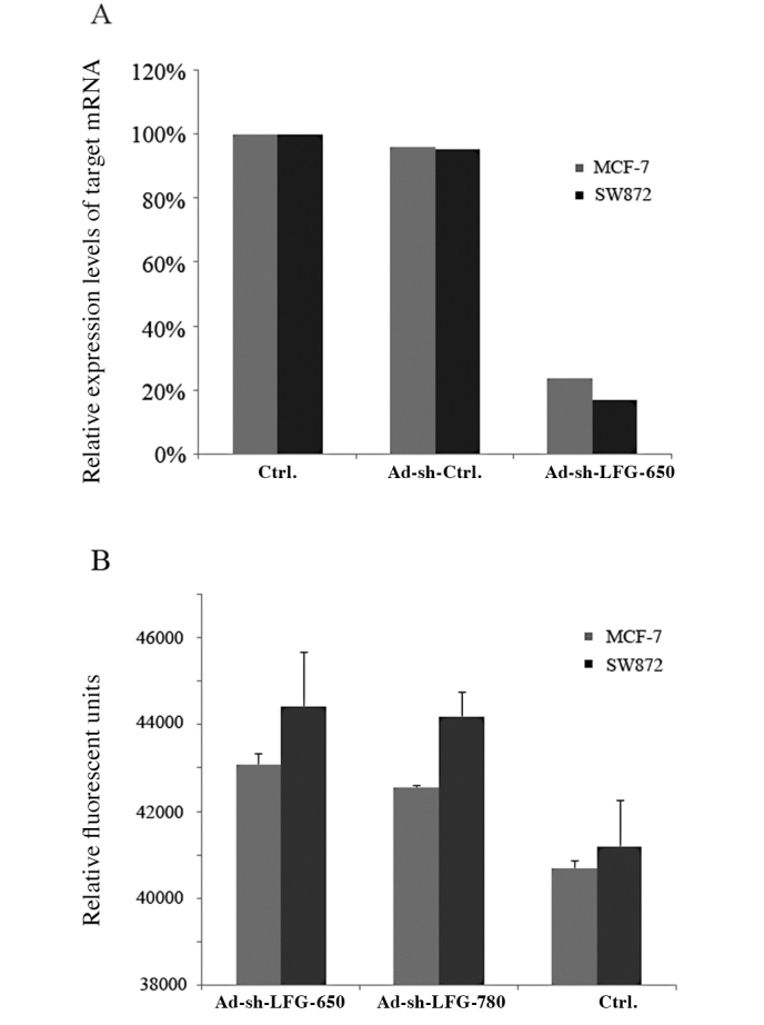
Figure 1.

(A) Reduction in LFG mRNA 48 h after transfection with adenoviral vector as compared to the control cells. Semi-quantitative RT-PCR was carried out in 20-μl samples with 5 ng cDNA and 10 pmol of each forward and reverse primer using the 2X SYBR-Green Sensi-Mix DNA kit. The relative gene expression was determined from the fluorescence intensity ratio of the target gene to 18S. (B) The human breast carcinoma MCF-7 and liposarcoma SW872 cell lines were transfected with Ad-sh-LFG-650 and Ad-sh-LFG-780 vectors for 48 h and analyzed for activated levels of caspase 3 using the Apo-One assay. The data are the means ± SD for triplicate determinations which were repeated in three separate experiments.
